Overview
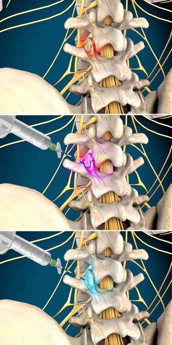
This diagnostic procedure is performed to identify a painful facet joint. The facet joints are the joints between the vertebrae in the spine. They allow the spine to bend, flex and twist.
Preparation
In preparation for the procedure, the patient is positioned on his stomach. The physician injects a local anesthetic. This numbs the skin and tissue around the facet joint that is suspected of causing the patient’s pain.
Contrast Dye Injected
Once this tissue is numb, the physician inserts a needle into the skin. The needle is carefully guided down to the facet joint. The physician injects a contrast solution through this needle. The contrast solution helps the physician see the area on a camera called a fluoroscope. The fluoroscope provides live x-ray images. The physician uses the fluoroscope to confirm the location of the needle’s tip.
Anesthetic Injected
Once the physician has confirmed that the needle is positioned correctly, the physician attaches a syringe containing an anesthetic medication. This medication is injected around small nerves called the medial branch nerves. These carry signals to and from the facet joints. The anesthetic will temporarily block sensation in these nerves.
End of Procedure
If the temporary injection relieves the patient’s pain, the physician may inject a more long-lasting anesthetic. If the temporary injection does not relieve the pain, the physician may test nearby facet joints to identify the correct one.

